Skip to content
Ákos Kappel
Language: en
Theme
Light
Dark

Glaucoma Segmentation

Master's thesis: neural networks that outline the optic disc and cup in eye images.
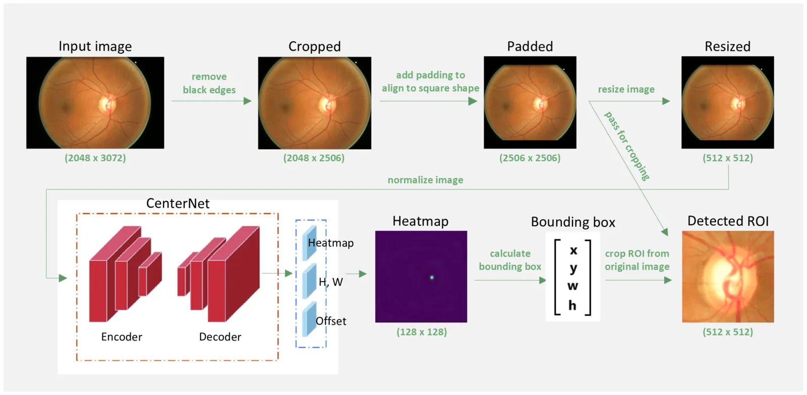
Processing steps from a raw eye image to the detected optic disc

About the project

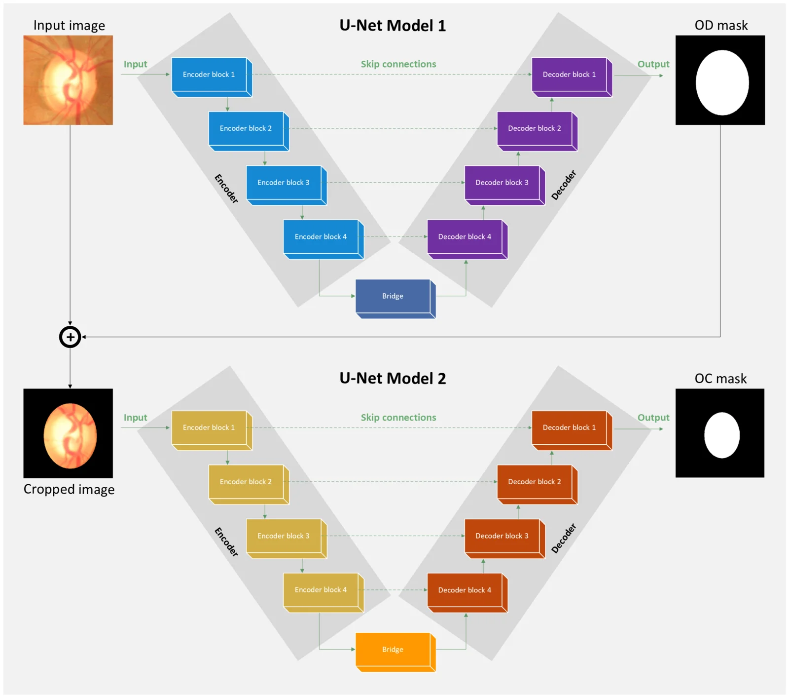
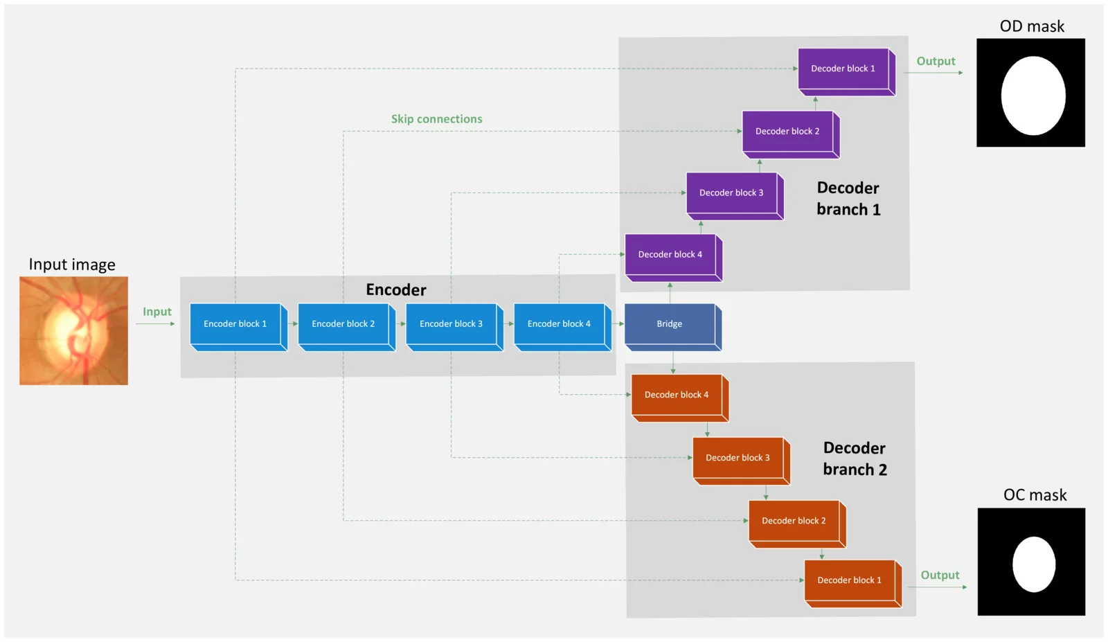
Glaucoma is diagnosed partly from the ratio between the optic cup and the optic disc. For my master's thesis I designed, implemented and evaluated two network architectures for finding them: a cascade, where a second network looks for the cup inside the disc found by the first, and a dual-decoder network with one shared encoder.

The models were trained on the ORIGA dataset and tested on DRISHTI-GS. The best one reached a Dice score of 96.8 % for the disc and 89.7 % for the cup.

Highlights

  • Cascade and dual-decoder architectures in PyTorch
  • Region-of-interest detection and training in polar coordinates
  • Explainability with Grad-CAM

Under the hood

  • Exploratory data analysis showed class imbalance and very different image quality, which shaped the preprocessing
  • A CenterNet model first finds the region of interest around the optic disc, and the segmentation runs on that crop in polar coordinates
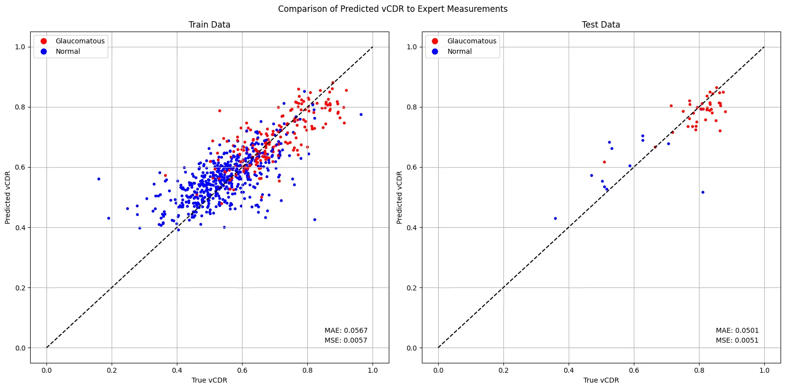
  • The vertical cup-to-disc ratio from the predictions was compared with values measured by experts
  • Probability maps, activation maps and Grad-CAM show what the networks look at
When
2023 – 2024
Type
University
Supervision
Supervised by doc. RNDr. Silvester Czanner, PhD.
Versions
  • Python 3.10
  • PyTorch 2.0
Stack
  • Python
  • PyTorch
  • OpenCV
  • NumPy
  • pandas
  • scikit-learn
  • Albumentations
  • Matplotlib
  • Jupyter
  • Weights & Biases
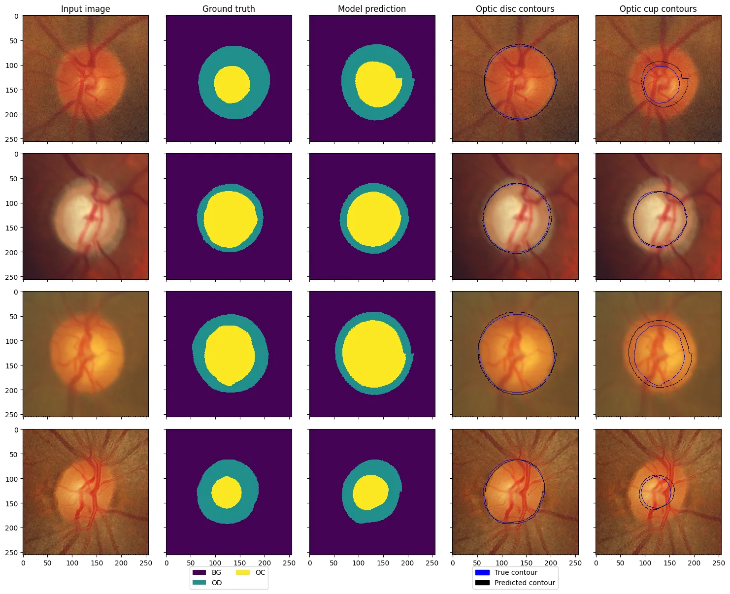
Four eye images with the expert outline, the model prediction and both contours overlaid
Results of the dual-decoder model: input, ground truth, prediction, and the true (blue) and predicted (black) contours of the optic disc and cup.
Diagram of two U-Net models in a row: the first outputs the disc mask, which crops the input for the second, which outputs the cup mask
Cascade architecture: a first U-Net segments the optic disc, its mask crops the image, and a second U-Net finds the cup inside it. It reached a Dice score of 96.8 % for the disc and 87.1 % for the cup.
Diagram of a network with one shared encoder and two decoders, one for the disc and one for the cup
Dual-decoder architecture: one shared encoder, a decoder for the optic disc and one for the cup. It reached a Dice score of 96.8 % for the disc and 89.7 % for the cup.
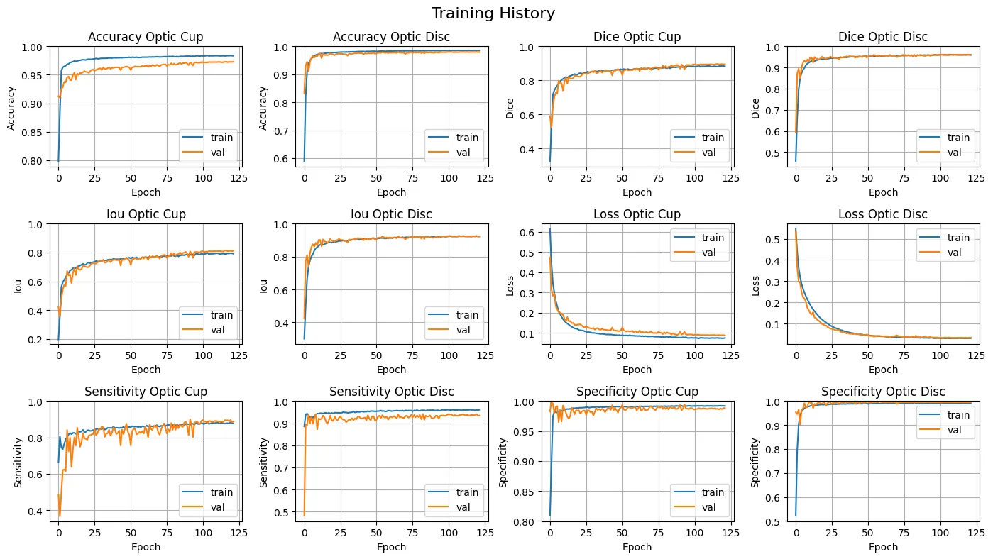
Line charts of loss and Dice score over the training epochs
Training history of the dual-decoder model: loss, Dice score and other metrics on the training and validation sets.
Charts comparing the predicted and the expert cup-to-disc ratio
Vertical cup-to-disc ratio, the main sign of glaucoma, computed from the predictions and compared with values measured by experts.

1 of 5

Four eye images with the expert outline, the model prediction and both contours overlaid

Results of the dual-decoder model: input, ground truth, prediction, and the true (blue) and predicted (black) contours of the optic disc and cup.